
In Clinical
Let’s get clinical. Follow the links below to find out more about the latest clinical insight in community pharmacy.Bookmark
It is generally accepted that an inflammatory skin condition, such as atopic eczema or allergic dermatitis, is characterised by erythema or redness due to increased blood flow to the affected area of skin. This conception is based on the notion that the individual is Caucasian.
Such conditions also occur in patients of different ethnicities but their non-white skin makes it more difficult to identify inflammatory changes. The term ‘skin of colour’ (SOC) is used to describe those with non-Caucasian skin and includes individuals of African, Latino, Asian and native Hawaiian descent.
In the past, the subject of SOC has been largely ignored but as the UK population becomes more culturally diverse, it is important to know how common skin conditions present in those with SOC and to have a better understanding of some of the conditions which are specific to those with darker skin tones.
Origin of different skin tones
The determination of an individual’s skin colour is based on the Fitzpatrick scale, which was developed largely to estimate the skin’s response to UV radiation.
The skin is divided into six types. Protection against UV radiation is provided by the pigment melanin, produced by melanocyte cells in the basal layer of the epidermis. Once formed, melanin is ‘packaged’ into intracellular organelles termed melanosomes. Melanocytes produce two forms of melanin: eumelanin, which is black-brown in colour, and pheomelanin, which is a red-yellow colour. Individuals of African descent produce a higher amount of eumelanin.
| Table 1: Fitzpatrick skin types | ||
| Skin type | Typical features | Tanning ability |
| 1 | Pale white skin, blue or green eyes/blonde or red hair | Always burns in the sun and doesn't tan |
| 2 | Fair skinned, blue eyes | Easily burns and poorly tans |
| 3 | Darker, white skin | Will tan after an initial burn |
| 4 | Light brown skin | Skin minimally or rarely burns and easily tans |
| 5 | Brown skin | Skin rarely burns, always tans darkly |
| 6 | Dark brown or black skin | Never burns and always tans darkly |
Contrary to popular belief, all individuals irrespective of their skin tone have a similar number of melanocyte cells. The reason for the observed differences in skin colour arises due to the size, distribution and type of melanin within the melanosomes. Thus, an individual of African descent has larger melanosomes with more eumelanin, which are distributed throughout the epidermis and not just in the basal layer as with Caucasians, making the overall appearance of the skin darker.
Common skin conditions in darker skin tones
Atopic eczema (AE) is a chronic, relapsing-remitting condition with prevalence data suggesting that African-American children are 1.7 times more likely to develop AE compared to European American children (Exp Dermatol, vol 4, 2018). The clinical presentation is also different.
In Caucasians, the classic presentation is of highly pruritic, erythematous lesions on flexor surfaces such as the inner elbow crease and behind the knees. In contrast, among children of African descent, the disease can occur on extensor surfaces, but a further distinguishing feature is the follicular or papular appearance. Additionally, the associated erythema presents as a slightly purple hue.
Some evidence also points to Black skin having as much as a 50 per cent lower ceramide content compared to Caucasian skin, which makes the skin prone to drying and dehydration and might explain why many Black individuals often use lots of emollients. In addition, among those with Asian skin, lesions tend to be more demarcated and with greater scaling and lichenification (i.e. skin thickening) and the associated erythema is a reddish-brown colour or even a purple or violaceous grey.
Fortunately, the management of AE in people with SOC is no different to Caucasians and involves the use of regular emollients and topical steroids to help manage disease flares.
Psoriasis in patients with darker skin is easily visible and has the same scaling appearance as in Caucasian skin. One potential differential for psoriasis that pharmacy teams need to be aware of is hypertrophic lichen planus, which is pruritic and can present on the shins and ankles.
If pharmacists are unsure of the diagnosis, they should refer patients to their GP. As with eczema, treatment of psoriasis in people with SOC is the same as for Caucasian skin.
One of the most common complications experienced by patients with SOC who have acne is the development of post-inflammatory hyperpigmentation (PIH). Exactly why this occurs remains to be determined but may be related to acne-induced changes in the processes regulating melanogenesis. In some cases, the degree of PIH is related to the severity of the acne.
Treatment of acne in those with SOC is generally the same, although some clinicians prescribe azelaic acid to help reduce PIH despite this being an unlicensed indication. In other instances, hydroquinone, which is a recognised skin lightening agent, has also been used.
A further problem for those with SOC is how topical agents such as retinoids or benzoyl peroxide, which typically cause dryness and irritation, can lead to increased PIH. The Primary Care Dermatology Society (PCDS) advocates that such topical treatments should be used less often. It suggests applying a topical agent in the evening and washing off before going to bed or even using every second or third day. In fact, the PCDS recommends that early and more aggressive treatment is required for those with SOC and this may even include referral for isotretinoin.
Finally, it is worth emphasising to patients that PIH can last for several months or even a year before clearing.
Acne keloidalis nuchae is a long-term, progressive chronic folliculitis which develops into keloid-like papules on the occipital region of the scalp. Treatment options are generally not very effective and topical antimicrobial agents are often used to prevent secondary infection.
If treated at an early stage, the condition can respond to potent topical steroids although intra-lesional injection of triamcinolone can sometimes help reduce the size of the lesions.
This is an extremely common inflammatory condition in the skin on the face and neck in people with SOC, particularly men of African descent due to their tight curly hair, and is commonly referred to as a shaving rash. While generally more common in men, the condition can occur in women who shave in the axillae and suprapubic regions.
When the skin is shaved, the cut hairs penetrate the skin and produce what is known as a foreign-body inflammatory reaction. The papules and pustules are generally sterile (i.e. not infected) although a secondary bacterial infection may develop.
Since an individual’s hairs are curly, the cut hair penetrates back into the skin and the condition only really completely resolves after hair removal. It is recommended that individuals avoid a very close shave or use clippers or even refrain from shaving every day.
Patient history
Inflammatory skin conditions such as atopic eczema affect those with skin of colour although clearly the associated inflammatory changes will not present with redness as in Caucasian skin.
As with any patient, a complete patient history should be taken, enquiring about family history and potential triggers, which are usually the same as for Caucasians.
Post-inflammatory hyperpigmentation represents a major concern for some patients with skin of colour and it is important to explain that the skin changes can take many months to resolve. Skin care routines should also be gentler given how skin of colour is prone to dryness and irritation.
With an increasing amount of support available (see ‘Useful resources’ box), community pharmacists should be able to provide the necessary help and counselling to people with skin of colour, ensuring equality of care which will hopefully reduce both diagnostic delays and a deterioration of their skin condition.
Diagnostic difficulties in SOC
Many of the diagnostic difficulties experienced by healthcare professionals in this area are likely to reflect inadequacies in their training, particularly given how dermatological diagnoses are visual and largely based on pattern recognition.
In a 2021 study published in the Journal of the American Academy of Dermatology (JAAD, vol 84, issue 1, 2021), which analysed the image content of six commonly used dermatology textbooks, it was shown that only 4-19 per cent of images showed dark skin.
Even for extremely common conditions such as acne, the researchers found that from a total of 69 images used in the textbooks, only three provided illustrative examples in people with darker skin.
This lack of imagery for people with SOC has also encroached on the diagnosis of Covid-19-related skin problems. In a study of an international registry of 716 patients from 31 countries (JAAD, vol 83, page 1118, 2020), only 1.9 per cent of all images were from those of Black skin and 5 per cent from Hispanic/Latino patients.
Useful resources
- Brown Skin Matters – this contains a huge image library with examples of how common skin conditions present in patients of colour
- Skin of Color Society – contains a similar wide range of resources
- British Association of Dermatologists – provides extensive education resources for healthcare professionals
Sponsored
 Sponsored education
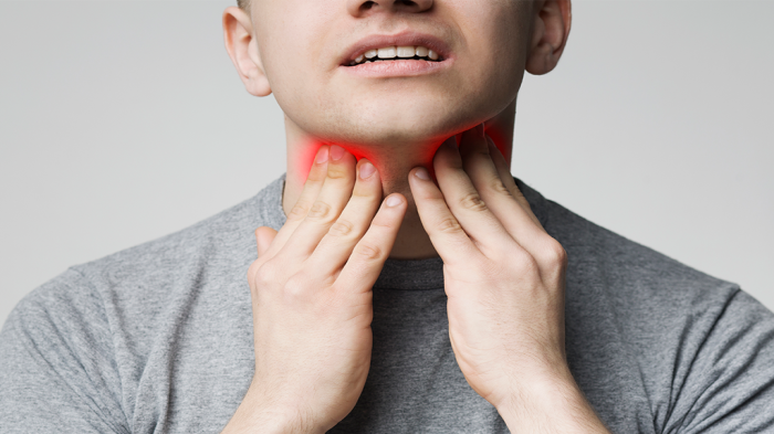
7 steps to managing sore throat
Get to grips with what customers want from their sore throat treatment and upgrade your consultations with this 7-step guide
 Sponsored education
Challenge your thinking on warts and verrucas
Discover different treatment options for warts and verruas and when to recommend them to your customers, based on their individual needs
Record my learning outcomes