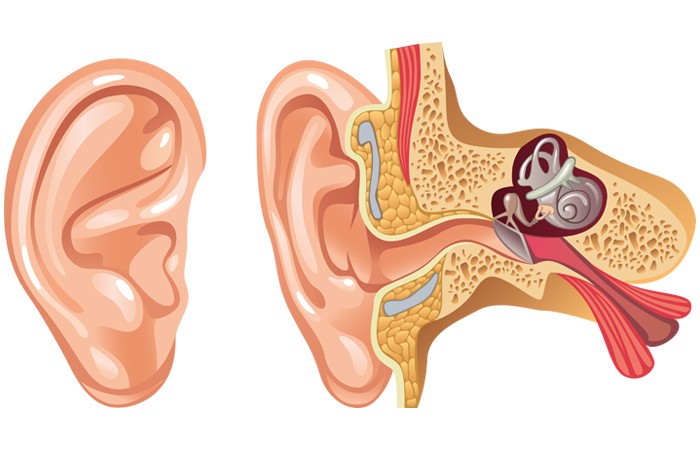
The anatomy of the ear
The ear has three main parts: the outer ear, which is the part that can be seen, the middle ear and the inner ear.

Breaking it down
The outer ear consists of the pinna (auricle), which is made of cartilage and skin, and the ear canal. The ear canal is around 2.5cm long and runs from the outer ear to the eardrum (tympanic membrane), which divides the outer and middle ear. Earwax is secreted in the ear canal, and acts as a defence against bacteria and particles in the air reaching the eardrum. External noise causes the eardrum to vibrate, transmitting sound to the middle ear.
The middle ear is an air-filled space behind the eardrum that contains tiny bones that move with the eardrum’s vibrations. These bones are called the malleus, incus and stapes (hammer, anvil and stirrup) – collectively known as the ossicles and are the smallest bones in the body. The stapes is the smallest of the three at around 3mm x 2.5mm. The Eustachian tube is also in the middle ear. It is connected to the throat via the back of the nose and has a role in making the pressure on both sides of the eardrum equal. It also drains fluid from the middle ear.
The inner ear (labyrinth) contains the spiral-shaped cochlea, which transforms sound waves into electrical impulses and sends them to the brain via the cochlear nerve, and the vestibular system. This consists of the vestibule and three semi-circular canals and is a complex system of fluid-filled passages that help maintain the body’s balance.
Sponsored
 Sponsored education
Challenge your thinking on warts and verrucas
Discover different treatment options for warts and verruas and when to recommend them to your customers, based on their individual needs
 Sponsored education
A different approach to pain
Complete this interactive video to rethink your pain recommendations and ensure you offer every customer the most appropriate advice
